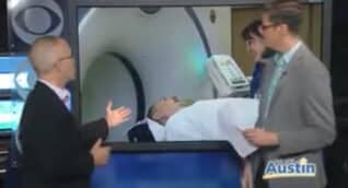
Molecular radiology is a specialty that uses compounds called radiopharmaceuticals to detect and treat disease. Radiopharmaceuticals are made by attaching a specialized biological compound to a radioactive molecule called a radionuclide. Depending on the type of compound, the radiopharmaceutical will go to different tissues in your body, such as the liver, bones, kidneys, thyroid, etc. The radiopharmaceutical is taken up by injured or diseased tissue in different amounts than in surrounding healthy tissue. This difference can be detected on specialized scanners or can be used to treat disease.
The Society of Nuclear Medicine and Molecular Imaging (SNMMI) has created a video on the value of Molecular Radiology. The video below highlights several specific examples—with corresponding evidence—of how nuclear radiology and molecular imaging are helping patients with cancer, heart disease, and other diseases.
The Value of Nuclear Medicine and Molecular Imaging
In a molecular radiology study, each radiopharmaceutical is targeted to a specific organ, such as bone or liver. The radiopharmaceutical may be injected, swallowed, or inhaled as a gas. A special scanner is then used to image body areas containing the radiopharmaceutical. The images produced by the scanner are interpreted by a radiologist who specializes in interpreting molecular radiology studies.
Yes. The radiation doses used in molecular radiology are similar to that of other imaging examinations and have been proven safe and effective over many decades of use. The molecular radiologist and technologist are trained in radiation safety procedures. All radiopharmaceuticals are manufactured under strict government standards and are administered in the smallest possible amounts needed to achieve quality imaging results. The radioactivity is quickly eliminated from your body usually within 24 hours after the test is completed. The risk of allergic reaction to the radiopharmaceutical is very low; no more than 2-3 reactions per 100,000 studies.
Pregnant women should be careful about imaging studies that use ionizing radiation, including X-ray, CT, and molecular radiology studies. Even though the radiation levels are low, there may still be a risk to the developing baby. Only in cases where the benefit clearly outweighs the risk should pregnant women undergo molecular radiology exams.
The radiopharmaceuticals used in these studies may be excreted in breast milk and could put nursing infants at risk. Therefore a breastfeeding woman should stop nursing for a period of time following a Molecular Radiology exam to allow the radiopharmaceutical to clear from her body. The amount of time needed to clear from the body varies for each specific radiopharmaceutical, lasting anywhere from a few hours to a few days. During that period, the woman is advised to express and discard her breastmilk. Once the radiopharmaceutical clears from the system, returning to breastfeeding is safe. The technologist or radiologist will provide information regarding the amount of time needed for the specific radiopharmaceutical that was given.
Bone & Joint Imaging
Cisternogram
DaTscan
Dementia Imaging
Gallium Scan
Gastric Emptying Scan
Gastrointestinal Bleeding Scan
Heat-Damaged Red Blood Cell Scan
Hemangioma Scan
HIDA Scan
Liver-Spleen Scan
Lymphoscintigraphy – lymphatic drainage
Lymphoscintigraphy – Sentinel Node
Meckel’s Diverticulum Scan
MIBG scan
Octreotide Scan
Parathyroid Scan
PET/CT
Radioiodine Thyroid Scan
Radioiodine Whole Body Scan
Radioiodine Therapy (I-131)
Radionuclide Ventriculogram (MUGA)
Renal Scan
SPECT/CT
White Blood Cell Scan

 Back to Top