About Ultrasound at ARA
Ultrasound is a medical imaging technique that uses high-frequency sound waves to produce images of the body’s internal structures. By using a special ultrasound transducer (probe), this test visualizes structures without exposing the patient to ionizing radiation (X-rays). Ultrasound has no known risks, and the procedure is safe for monitoring pregnant women and developing infants during pregnancy.
Ultrasound is a fast, non-invasive method for doctors to evaluate soft tissues that do not show up on regular X-ray exams, including mammography. This technique is especially useful to visualize the appearance, size, consistency, and shape of internal organs and abnormalities. Ultrasound can be used to examine organs and detect tumors, infections, or fluid-filled structures. The images can also be visualized in real-time on a monitor.
Ultrasound Exams
Choose a speciality from the list below to learn more about a specific service.
While ultrasound images may look mysterious to the untrained eye, to ARA's experienced radiologists and technologists, they can be the key to discovering something important about your body.
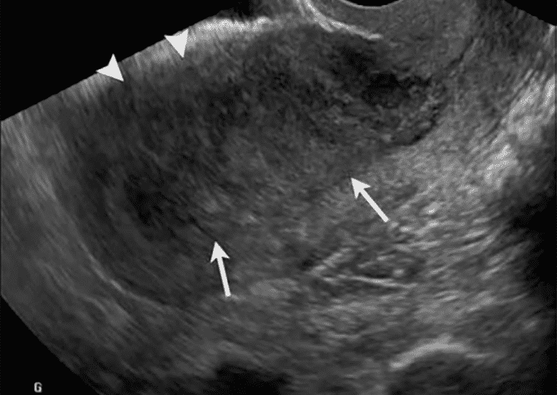
This ultrasound is an image of a uterus with a thickened endometrium, which is an important factor in diagnosing uterine cancer.
Ultrasound images are created by sending sound waves through a specialized wand which also records the signals that are bounced back by structures in the body.
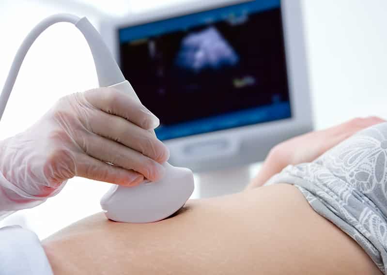
The ultrasound wand glides easily over lubricated skin as the technologist makes sure the right pictures are obtained for the radiologist to review and make a diagnosis.
How does ultrasound work?
The ultrasound transducer sends out small pulses of high-frequency sound waves. When pressed against the skin, the transducer transmits sound waves that bounce off structures in the body. The transducer picks up the rebounding sound waves, and, with the help of a computer, the characteristics of a structure can be determined.
Ultrasound can be used to help guide doctors when taking tissue biopsies using a technique called ultrasound-guided biopsy. Doppler ultrasound is an advanced ultrasound technique that uses a computer to measure blood flow speed and direction.
 Back to Top