Radiation Safety
Radiation risks in perspective.
At ARA, your safety is our mission.
As a patient, you may have concerns about the imaging procedures prescribed for you. The medical experts at ARA take the use of radiation very seriously. We strongly believe that when we do an exam that uses radiation, the benefits must far outweigh the risk. We are dedicated to our branch of medicine – imaging that allows us to see inside the body in ways that avoid harm to the patient, such as exploratory surgery and other damaging procedures that imaging makes unnecessary.
ARA wants you to be informed about the steps our practice takes to ensure these risks are limited, the many types of imaging that do/do not not use radiation, the risks of exposure, what you can do to help, and where you can go for further information.
Our focus is to limit your radiation exposure
- ARA has over 115 board-certified radiologists, all of whom have extensive training in radiation safety and methods to limit radiation exposure for our patients. One physician serves as Radiation Safety Officer to provide leadership and set standards for our radiation safety programs. All ARA staff and physicians adhere to the “as low as reasonably achievable” standard in keeping radiation doses low while producing scans that are useful for diagnostics and procedures.
- All of our technical staff have national and state registration and certifications and are trained to monitor the radiation exposure of the equipment they use to perform your imaging.
- The imaging equipment at all of our facilities is American College of Radiology (ACR) accredited. As a requirement of this accreditation, each unit is calibrated and monitored by our staff and a licensed medical physicist and serviced by factory-qualified engineers. Buying and maintaining up-to-date equipment allows ARA to continue to keep your radiation dose as low as possible.
- Our practice supports The Alliance for Radiation Safety in Pediatric Imaging and the Image Gently® Campaign to lower radiation doses in the imaging of children. We also support the Image Wisely awareness program to encourage practitioners to avoid unnecessary scans and use the lowest optimal dose for necessary studies.
- Our radiologists can confer with your physician to ensure the most appropriate imaging procedure is performed to avoid unnecessary radiation exposure.
- Through our affiliation with the American College of Radiology and the American Board of Radiology, we monitor the latest publications and trends so that we may quickly implement any new dose reduction guidelines.
Understanding radiation in breast imaging
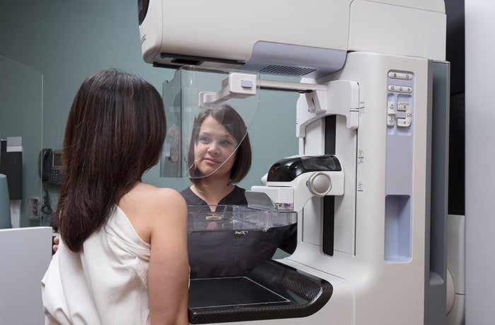
According to the American College of Radiology, the total dose for a typical mammogram with two views of each breast is 0.4 millisieverts (mSv), and the total dose for bone densitometry is 0.001 mSv. To help put those numbers into perspective, people are exposed to about 2 mSv of radiation each year from our natural surroundings. This would mean that getting a mammogram is the equivalent of only seven weeks in our normal, everyday life.
0.001
Bone densitometry exam radiation dose in mSv
0.4
Mammography exam radiation dose in mSv
50
Maximum annual limit of radiation dose in mSv for healthcare workers

ARA strongly believes that when we do an exam that uses radiation, the benefits must far outweigh the risk.
All types of imaging
Listed below are some common types of imaging, the radiation risk associated with each, and an example of comparable risk. Each exam within a modality will vary in its radiation dose based on the body area examined, such as the chest, abdomen, or extremity.
- Magnetic resonance imaging (MRI) does not use radiation during the examination. MRI uses magnetic fields and radio-frequency waves to image the body. There is no radiation risk involved with MRI.
- Ultrasound (US) exams do not use radiation during the examination. Ultrasound uses sound waves to image the body. There is no radiation risk involved with ultrasound.
- X-ray uses a minimal dose of radiation during the examination.
- Mammography uses a minimal dose of radiation during the examination. A mammogram examination is equivalent to approximately three months of background radiation. Note: Concern is sometimes raised about the effects of mammography on the thyroid and the use of a thyroid shield during a mammogram. Experts do not recommend the use of a thyroid shield because the amount of radiation received by the thyroid during a mammogram is extremely low, and the use of the shield can cause shadows and artifacts on the mammogram, necessitating further mammograms.
- Computed tomography (CT) uses more radiation than plain X-ray because it produces a more detailed image. The diagnostic benefit usually outweighs the radiation risk, so patients and their referring physicians should consider these risks and benefits. ARA’s CTs are a new generation of low-dose scanners that deliver superior images with less radiation.
- Fluoroscopy uses more radiation than plain X-ray because it uses an X-ray beam that passes continuously through the body to create a moving image. The image is projected on a monitor, allowing radiologists to see internal organs’ movement in real-time. The diagnostic benefit usually outweighs the radiation risk, so patients and their referring healthcare providers should consider these risks and benefits.
- Molecular radiology uses small amounts of radioactive materials (radiotracers), which are either injected or swallowed to target certain organs or areas of the body for imaging. The majority of the radiation received during treatment is excreted naturally from the body.
- Positron emission tomography (PET) uses small amounts of radioactive materials (radiotracers), which are injected to image the body, typically to help diagnose or stage cancer in a patient.
Understanding radiation in our daily lives
Each of us is exposed to radiation every day of our lives. According to the Environmental Protection Agency, the average person in the United States receives a dose of about 360 millirems (used to measure radiation) per year. 80% of that comes from natural sources such as radon gas, outer space (cosmic radiation), soil, and rocks. The remaining 20% comes from man-made radiation sources, primarily medical imaging. As an example, the typical chest X-ray is equivalent to the amount of radiation one experiences from our natural surroundings in approximately ten days.
It’s important to remember that negative effects from radiation are extremely rare. Only a small percentage of people who are heavily exposed to radiation develop radiation-induced cancer later in life. This includes people who are exposed to radiation from nuclear weapons, are involved in radiation accidents, and receive high-dose radiation therapy for cancer.
How to limit your radiation exposure
- Track your radiation exposure.
As part of your medical history, keep a list of imaging procedures you have undergone, including type, date, and locations. ARA will have a history of any imaging done at our facilities. - Discuss the exam with your physician.
Why do I need this exam? How will having this exam improve my healthcare? Are there alternatives that do not use radiation that will provide the same exam quality? - Ask questions.
Is the facility providing my imaging accredited by an official organization such as the American College of Radiology (ACR)? Are the technologists certified? Are the physicians reading my exam sub-specialized, board-certified radiologists?
More information
radiologyinfo.org Click on the safety link. Presented by the American College of Radiology (ACR) and the Radiological Society of North America (RSNA).
radiationanswers.org Presented by the Health Physics Society.
imagegently.org Image Gently® – see the parent section for pediatric imaging and what you can do.
imagewisely.org Presented by the American College of Radiology (ACR), the Radiological Society of North America (RSNA), the American Association of Physicists in Medicine (AAPM), and the American Society of Radiologic Technologists (ASRT).
epa.gov/radiation Environmental Protection Agency.
 Back to Top