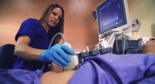
Vascular ultrasound is an imaging technique that uses sound waves to evaluate the body’s circulatory system (larger arteries and veins). This exam can be used to detect blockages in circulation, blood clots, vascular malformations, and other disorders associated with circulation. By using a special ultrasound probe, this test is performed in a non-invasive way and without exposing you to ionizing radiation (X-rays).
Doppler ultrasound is a special ultrasound technique that can measure blood flow speed and direction and is usually a part of vascular ultrasound exams.
More basic information on ultrasound is available in the About Ultrasound section.
Vascular ultrasound may be used in situations when symptoms suggest a problem with circulation. The test is convenient and non-invasive, and it gives doctors valuable diagnostic information quickly. Some situations where vascular ultrasound may be recommended include:
- Evaluating blood flow to body organs.
- Diagnosing blockages, plaques, or emboli.
- Detecting blood clots (deep vein thrombosis or DVT) in large veins.
- Evaluating patients in preparation for angioplasty.
- Assessing blood flow in blood vessel grafts and bypasses.
- Detecting aneurysm (abnormal artery enlargement).
- Evaluating varicose veins.
- Detecting abnormal blood flow due to tumors, vascular malformations, or infection.
Benefits
- Vascular ultrasound is an inexpensive, fast, and non-invasive way to assess circulation abnormalities including blood flow problems. Ultrasound may prevent the need to perform more invasive studies or procedures.
- Ultrasound does not expose you to any ionizing radiation (X-rays).
- Unlike MRI, ultrasound can be used in patients with any type of metal in their body, including implantable medical devices.
- Ultrasound is very safe and has no side effects.
- There is no need to administer contrast for ultrasound exams.
- Ultrasound can detect soft tissue abnormalities that cannot be visualized with regular X-ray exams.
Risks
- The use of diagnostic ultrasound has no known risks or harmful effects.
- Vascular ultrasound is done in an ARA imaging center. The exam will take about 30 minutes and you will be in the imaging center for about an hour.
- You may be asked to change into a gown.
- You will be placed lying on your back on an exam table. You may have to change position during the exam.
- A water-based gel will be placed on the skin over the area to be examined. The gel creates a sealed contact between your skin and the ultrasound probe. This eliminates any air pockets that may interfere with imaging. The probe will be moved around to capture images from different locations. The gel does not stain clothing.
- The technologist or doctor performing the study may have to apply pressure with the probe to the body part being examined. If the area is tender, you may experience some discomfort.
- You can return to your normal activities after the exam is over.
- Wear comfortable, loose-fitting clothing for the exam.
- If the abdominal area is being examined, and it is not an emergency, you may be asked to fast overnight before the exam.
To schedule a vascular ultrasound, please use our online scheduling tool in the Patient Portal or you may call our scheduling team at (512) 453-6100 or toll free at (800) 998-8214. A referral from your healthcare provider is required to make an appointment.
A radiologist, a physician specifically trained to interpret radiological examinations, will analyze the images and send a signed report to the provider who referred you to ARA. The physician will then share the results with you.

 Back to Top