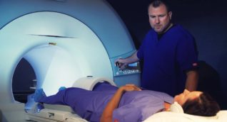
Magnetic resonance imaging (MRI) of the breast is a noninvasive test that is recommended to find and diagnose breast disease. MRI uses a powerful magnetic field, radio frequency pulses, and a computer to produce detailed images of body structure. These images are viewed on a computer monitor and can be transmitted electronically. MRI does not use ionizing radiation (X-rays). Radiologists use these detailed images to evaluate the features of the breast. MRI of the breast offers valuable information about many breast conditions that other imaging modalities cannot obtain. Breast MRI uses an intravenous contrast agent that increases the visibility of breast cancer on the MRI image.
- Screening for women at high risk of breast cancer. MRI may be the right screening tool for women with a strong family history or genetic predisposition to breast cancer. A strong family history might include a parent, sibling, or child who has had breast cancer before age 50 or close relatives that have had ovarian cancer. ARA includes a screening questionnaire on your intake form that can help you and your provider determine your risk. ARA generally recommends that women at high risk for breast cancer have an annual screening mammogram and an annual breast MRI scheduled six months apart.
- Screening women with dense breast tissue for breast cancer. Unlike mammography, MRI imaging is not affected by dense breast tissue, making it very effective in finding cancer in dense breasts.
- Evaluating a new diagnosis of breast cancer. MRI can be used to determine the size and scope of the breast cancer, if there are other abnormalities in the same breast or in the other breast, and if there is enlargement of the lymph nodes that may indicate the spread of cancer.
- Evaluating chemotherapy response before surgery. In some cases, breast cancer will be treated with chemotherapy before it is removed surgically. This is called neoadjuvant chemotherapy. MRI is used to monitor the effect of chemotherapy on the tumor.
- Evaluating breast implants. MRI is used to see if there has been a change or rupture in a breast implant.
Since MRI uses a very strong magnet, any implanted medical device may be affected, so be sure to tell your scheduler and technologist about any device in your body. In general, metallic orthopedic implants are not affected by MRI, but tell your radiologist about any implant you have before scheduling the exam. Your implant or device may come with a special information card that you should show to the radiology technician.
Some implants are not compatible with MRI scanners. Do not enter any MRI scanning area if you have any of the following implants:
- Cochlear (ear) implant
- Some brain aneurysm clips
- Some metal coils/stents placed inside blood vessels
- Cardiac defibrillators and pacemakers
Other implants that should be brought to the technologist’s attention before entering the MRI scanning area are:
- Artificial heart valves
- Implanted drug infusion ports
- Artificial limbs or metallic joint prostheses
- Implanted nerve stimulators
- Metal pins, screws, plates, stents, or surgical staples
Also, you should notify your doctor or technologist if you have any other metal in your body (shrapnel, bullets, needles, etc.). Metal in or near the eye is especially dangerous since any movement of the metal during the procedure could lead to eye damage. Dental fillings and braces usually are not affected by the magnetic field, but they may distort images taken of the head or face.
Benefits
- As a supplement to mammography, MRI has been proven to be effective in evaluating women at high risk for breast cancer.
- MRI can be used as an imaging guide for a minimally invasive needle biopsy for abnormalities identified only on MRI.
- MRI is an excellent imaging exam for women with dense breast tissue and for women with breast implants.
- MRI is useful in evaluating and staging breast cancer.
- Most people well tolerate the contrast agent used for MRI exams.
- MRI is a noninvasive and painless imaging technique that does not use ionizing radiation (X-rays).
Risks
- There is almost no risk to the average person getting an MRI exam when appropriate safety guidelines are observed.
- Because a very strong magnet is used, any metal implants or objects in the body may malfunction or cause a poor result on the MRI image. In these cases, it may not be possible for the patient to undergo an MRI exam. Precaution must be taken to remove all metal jewelry, watches, or body braces before the exam, including metal zippers and buttons on clothing.
- Patients with poor kidney function may be at risk for a rare complication thought to be caused by injections of high doses of a gadolinium-based contrast agent. Therefore, kidney function is assessed before using a contrast injection. For more information, please see About MRI Contrast.
- Very rarely, a patient will have an allergic reaction to the contrast material. These reactions are generally very mild and easily controlled with medication. A radiologist or paramedic will be available for immediate assistance if you have an allergic reaction during the exam.
- The American College of Radiology notes that available data suggest that it is safe to continue breastfeeding after receiving contrast. Please discuss your breastfeeding options with your medical provider.
- MRI produces images of the body’s structure by passing radio waves through a powerful magnetic field. Ionizing radiation (X-rays) is not used for this exam.
- You will be asked to change into a gown.
- Since you will be positioned within a large, strong magnet, you must remove all metal objects, including piercings.
- Let your technologist know if you have any medical or electronic implants or tattoos on your body.
- Breast MRI requires the injection of a contrast material that will help highlight any suspicious areas. After an initial series of MRI images, you will receive an intravenous (IV) injection that will be administered into a vein in your arm or hand by an ARA technologist.
- During your exam, you will be positioned on a special MRI table adapter that will allow you to lie on your stomach with your breasts hanging freely through an opening so they are in the correct position for a successful scan. Your arms will be placed above your head.
- MRI scanners are constructed with short tunnels that are open on both ends. Most people do not find this to be uncomfortable, but if you are anxious, please mention this to your scheduler. ARA may be able to schedule your exam with light sedation. ARA technologists are experts at helping people through MRI exams with minimum anxiety.
- The table will move into the magnet, and you must remain still as the exam proceeds.
- You will hear various noises as the MRI does its job, ranging from buzzing to loud knocking sounds.
- Several sets of images will be taken from different angles.
- The technologist will inform you of the exam’s progress, and you can contact and speak with the technologist at any time.
- The imaging session will last between 30 minutes and one hour, and you will be at ARA for about an hour and a half.
- Breast MRI requires a provider referral. Please bring a copy to your appointment.
- If you have had prior breast imaging at an office other than ARA Diagnostic Imaging, please inform the scheduler or bring those images with you for comparison if possible.
- Let the scheduler and technologist know if you have medical or electronic devices in your body, have any kidney issues or allergies to contrast materials, or are pregnant or nursing.
- If you have mobility or pain issues, please let your scheduler know so we can make accommodations.
- If you are scheduled for sedation, please arrive earlier than your scheduled appointment time. All patients receiving sedation must have someone with them to drive them home after the procedure.
- If you feel anxious about claustrophobia during the exam, please discuss this with your scheduler. ARA can discuss options to help make you more comfortable, such as light sedation.
To schedule, please call our scheduling team at (512) 453-6100 or toll free at (800) 998-8214. A provider referral is required to make an appointment.
A radiologist, a physician specifically trained to interpret radiological examinations, will analyze the images and send a signed report to the provider who referred you to ARA. The physician will then share the results with you.
ARA wants to provide a safe, comfortable environment for patients and staff.
Patients may either bring or request a chaperone to accompany them during their exam to help protect and enhance their safety and comfort.
When requested, ARA will attempt to provide a chaperone with whom the patient feels comfortable. If a patient’s chaperone request cannot be accommodated, the patient will be given the opportunity to reschedule their exam.

