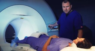
MR arthrography (arthrogram) is a contrast-enhanced study used to visualize the interior of joints in a very detailed way. An arthrogram helps doctors diagnose problems with the bones, cartilage, ligaments, and tendons within a joint. Arthrograms are especially useful for determining the cause of unexplained joint pain.
Contrast material (gadolinium based) is injected directly into the joint. The fluid expands the joint and provides a better view of internal joint structures. Joint injections are done immediately before the MRI exam using fluoroscopic guidance. Find out more about Fluoroscopy.
An arthrogram is recommended when a doctor, typically an orthopedic surgeon, requires visualization of the interior of a joint. For example, if you have a joint injury, an arthrogram might be recommended to determine the extent of injury. Arthrograms are used to evaluate problems in a variety of joints, such as the shoulder, elbow, wrist, hip, knee, and ankle.
Arthrograms are a diagnostic tool which may also determine whether you require surgery for a joint problem.
Benefits
- Arthrograms are a minimally invasive way for doctors to evaluate the interior of a joint. This could avoid the need for surgical exploration of the joint.
- Even if joint surgery is eventually required, MR arthrography gives surgeons more detailed information about the joint prior to surgery. This may improve the outcome or shorten the length of recovery.
- A variety of problems can be evaluated by MR arthrogram. For example, doctors can evaluate the shoulder for labral tears. Knees can be examined for damage to cartilage and ligaments.
- No ionizing radiation is used in MRI exams. If you get a gadolinium injection into the joint being imaged, fluoroscopy will be used to guide the needle to the proper spot. Fluoroscopy uses a low dose of radiation because it uses X-ray technology, but the benefit of an accurate diagnosis far outweighs the risk. Please refer to the section About Fluoroscopy for more information on the risk of radiation used in this exam.
- MR arthrography may be able to visualize some structures better than other joint imaging methods.
Risks
- Any medical devices implanted into your body may be at risk of malfunction due to the strong magnetic environment. See Can I have an MRI if I have metal in my body?
- In very rare cases, in patients with poor kidney function, nephrogenic systemic fibrosis is a possible complication when contrast is used. Please refer to the About MRI Contrast section for more details.
- Gadolinium-based contrast has a very slight risk of causing an allergic reaction which can usually be easily treated.
- No ionizing radiation is used in MRI exams. If you get a gadolinium injection into the joint being imaged, fluoroscopy will be used to guide the needle to the proper spot. Fluoroscopy uses a low dose of radiation because it uses X-ray technology, but the benefit of an accurate diagnosis far outweighs the risk. Please refer to the section About Fluoroscopy for more information on the risk of radiation used in this exam.
- Pregnant women should consult with their physician prior to an MRI exam. However, there have been no documented negative effects of MRI in the many years of medical usage of MRI, and MRI is often the method of imaging chosen for pregnant women and fetuses. It should be noted that MRI contrast agents are not recommended to be used during pregnancy unless the benefits far outweigh the risks.
- The ACR states that current information suggests breastfeeding is safe after the use of intravenous contrast. Please discuss your breastfeeding options with your medical provider.
- MR arthrogram is provided in multiple ARA imaging centers.
- You will be asked to change into a gown. You will be required to remove all metal objects from your body.
- You may have X-rays taken before the MRI to have a baseline exam to refer to later.
- If you are having a joint injection of contrast agent (direct arthrogram), your technologist will use fluoroscopy imaging to help guide the needle to the right spot in your joint. The contrast agent will expand the joint and enhance the detail on the MRI image.
- The area to be injected is cleansed with antiseptic and a sterile drape placed around the injection site. Using a small needle, your technologist will inject local anesthetic. Once the area is numb, a larger needle will be used to inject the contrast material. In some cases, joint fluid is removed with a needle prior to injection.
- Medication, such as steroids, may be injected into the joint during the exam for treatment purposes.
- Next, you will move to the MRI portion of your exam. You’ll be placed on a moveable exam table which may have straps or bolsters to help keep your body from moving.
- Special coils that enhance the MRI signal will be placed near the parts of the body to be examined.
- The technologist will leave the MRI room where you are and conduct the exam from a computer in a control room. You will be able to speak with the technologist at all times. The technologist has direct visualization of you and the room. They will keep you informed of what is happening as your exam progresses.
- When the procedure begins, the table moves you into the magnet, which is a cylinder-shaped machine.
- MRI scanners are constructed with short tunnels that are open on both ends. Most people do not find this to be uncomfortable, but if you are anxious, please mention this to your scheduler. ARA may be able to schedule your exam on one of our open or short-bore MRI scanners or plan for you to have light sedation. Our clinical staff are experts at helping people through MRI exams with minimum anxiety.
- During the exam you might feel warmth in the body part being examined. If this becomes uncomfortable, let your technologist know.
- When the images are being taken, you will hear loud knocking, tapping or thumping sounds. Earplugs or headphones with music are provided before the test begins.
- In rare cases, some patients experience side effects from the contrast material, such as nausea or headache. If you experience any discomfort, let the technologist know.
- You will be required to change into a gown. If possible, leave all jewelry and metal objects at home.
- Unless you are told otherwise, you may follow your normal diet and routine before the exam. You should take all your medications as usual.
- Some MRI exams require a prep which may include no food or drink for a specified time before the exam. Your scheduler will inform you of preparations if needed. If you are not given special instructions, you may follow your normal diet and routine before the exam.
- Ask your doctor for specific directions about your daily medications if you have been asked to refrain from taking them.
- Be sure to tell your technologist about any illness or allergies you may have. Also, provide a list of your current medications.
- Be sure to tell your scheduler and technologist if you are or might be pregnant.
- If you suffer from claustrophobia, panic attacks or anxiety, you might choose to receive sedation before the exam. Please mention this to your scheduler when making your appointment.
- Before you schedule an MRI, refer to the section Can I have an MRI if I have metal in my body? to get more information about metal devices, implants, or any other metal that might be affected by the magnet.
- In some cases, an X-ray may be taken before the MRI to detect any metal that could be located inside your body.
To schedule an MR arthrogram, please use our online scheduling tool in the Patient Portal or you may call our scheduling team at (512) 453-6100 or toll free at (800) 998-8214. A referral from your healthcare provider is required to make an appointment.
A radiologist, a physician specifically trained to interpret radiological examinations, will analyze the images and send a signed report to the provider who referred you to ARA. The physician will then share the results with you.

