CT Scan
Fine detail for a quick diagnosis.
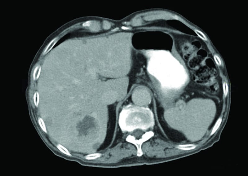
Computed tomography, commonly known as CT or CAT scan, uses X-rays and computer technology to generate multiple, detailed images of the internal organs, bones, soft tissue, and blood vessels, producing views inside the body in slices similar to a loaf of bread. Using advanced computer programming, some CT scan computers can generate 3D images. CT scans help doctors visualize the inside of the body to look for abnormalities, disease, tumors, infections, or anatomic malformations. Images are viewed on a computer screen and can be shared electronically which helps doctors diagnose problems at a distance.
60%
ARA’s latest clinic-wide upgrade of CT scanners allows for an up to 60% lower radiation dose than previous units.
CT Scan Services
Choose a speciality from the list below to learn more about a specific service.

A CT scan can produce images of your body in multiple slices, somewhat like the slices in a loaf of bread.
This allows radiologists to see the inside of your body in great detail.


CT Imaging is often used in emergency situations because it is a fast and detailed imaging modality that allows doctors to assess your condition quickly.
CT is also used to find cancer in your body and get images of tumors and their blood supply without needing to do exploratory surgery.

What is CT imaging used for?
CT imaging is often the best method for detecting many cancers since it allows radiologists to confirm the presence of a tumor and determine its size, shape, location, and what kind of blood vessels are supplying it, all without surgery. CT is also frequently used to assess internal injuries in emergency situations, helping health care providers respond quickly.
CT scans are fast, painless and non-invasive. Depending on the purpose of the exam and the needs of the patient, a CT scan may be performed with or without contrast.
During a CT scan, you are briefly exposed to ionizing radiation (X-rays). However, CT scans use low dose radiation and have not been shown to cause harm when deemed diagnostically appropriate. ARA’s latest clinic-wide upgrade of CT scanners allows for an up to 60% lower radiation dose than previous units.
How does a CT scan work?
With CT scanning, multiple X-ray beams and electronic X-ray detectors rotate around you as you lie on a table. The system measures the amount of radiation absorbed by your body, and a computer uses the information to generate images. CT scan images are cross-sectional which means they show a thin section, or slice, of the body in each image, somewhat like the slices in a loaf of bread.
What if I am pregnant? Can I still have a CT scan?
If you are, or think you are pregnant, be sure to notify your doctor or technologist before undergoing a CT scan. The amount of radiation received during a CT scan is unlikely to harm you or your baby. However, in general, CT scans are not recommended in pregnant women. In every case, the mother’s health must be considered as well. The benefit to the pregnant woman of having the CT scan to diagnose an illness may outweigh the small amount of risk to the baby from a low-dose CT scan.
The part of your body being scanned should also be considered. For example, brain CT exposes the unborn baby to little or no radiation. Even if the fetus is directly exposed to CT scan radiation (such as in CT scans of the abdomen or pelvis), the increased risk of developing cancer later in life is one in 1000. Some doctors may recommend another type of exam (ultrasound or MRI) to avoid exposing your baby to radiation.
 Back to Top