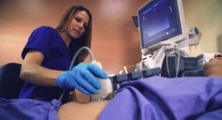
Ultrasound is a noninvasive, safe, and painless imaging tool that uses sound waves to produce pictures of the inside of the breast which are used to evaluate breast abnormalities. In ultrasound, also called sonography, a water-based gel is applied to the skin and a small, hand-held transducer (probe) is placed on the breast in the area to be examined. High-frequency sound waves are sent from the probe into the body and the probe collects sounds that bounce back and transmits them to a computer. As the technologist moves the transducer around the examination area, a computer creates an image that radiologists use to find disease.
- Evaluating a breast abnormality. Ultrasound is used to diagnose breast abnormalities after detection in a physical or imaging examination. It can help determine if a lump is solid in nature or fluid-filled (such as a benign cyst) or both.
- Supplemental breast cancer screening. While mammography is the gold standard for breast screening, mammograms do not detect all breast cancers. Ultrasound may be used as a supplemental screening for women with dense breast tissue (a lot of glandular and connective tissue as compared to fatty tissue).
- Ultrasound-guided breast biopsy. When a suspicious abnormality has been found in your breast, your radiologist and/or medical provider may recommend that a biopsy, or tissue sample, be taken. One way to sample tissue from the breast is to locate the abnormality using ultrasound and guide a hollow needle to the site to take a core biopsy. See Ultrasound-Guided Breast Biopsy for more information.
Benefits
- Ultrasound is a noninvasive imaging technique that does not use ionizing radiation (X-rays).
- Occasionally, ultrasound may require steady pressure on the body part being examined but it is generally just uncomfortable and not painful. Ultrasound is easily tolerated by most patients.
- Ultrasound is an excellent imaging exam for soft tissues.
- Ultrasound is real-time imaging which can be a good tool for guiding minimally invasive procedures such as needle biopsy.
- Ultrasound can help detect lesions in women with dense breasts.
Risks
- There are no known harmful effects from standard diagnostic ultrasound.
- It is possible that you will get a “false positive,” as breast ultrasound may indicate the need for more testing or biopsy. Most biopsies are done with a small needle and only a small percentage are positive for cancer.
- You will be asked to change into a gown and lie on your back or side on the examining table. Depending on the location of the area to be examined, you may be asked to raise your arm above your head.
- The technologist or radiologist will apply a warm, water-based gel to the area being studied. The gel helps the transducer (probe) make secure contact with the skin and eliminate air pockets that might block the sound waves and effect the image. The gel is easily wiped off and does not stain clothing.
- The technologist will move the transducer around the area, capturing images from different angles. There is usually no pain or discomfort, but you may feel pressure or minor pain if the scanner moves over tender areas of the breast.
- A radiologist experienced in ultrasound will view the images and may request further ultrasound images.
- Breast ultrasound usually takes about 30 minutes. You will be at ARA for about an hour.
- Breast ultrasound requires a provider referral. Please bring a copy to your appointment.
- Since you will undress from the waist up for the exam, you may wish to wear a two-piece outfit.
To schedule a breast ultrasound, please use our convenient online scheduling tool in the Patient Portal or you may call our scheduling team at (512) 453-6100 or toll free at (800) 998-8214. A referral from your healthcare provider is required to make an appointment.
A radiologist, a physician specifically trained to interpret radiological examinations, will analyze the images and send a signed report to the provider who referred you to ARA. The physician will then share the results with you.
ARA wants to provide a safe, comfortable environment for patients and staff.
Patients may either bring or request a chaperone to accompany them during their breast ultrasound to help protect and enhance their safety and comfort.
When requested, ARA will attempt to provide a chaperone with whom the patient feels comfortable. If a patient’s chaperone request cannot be accommodated, the patient will be given the opportunity to reschedule their exam.
 Back to Top